High-Content Cell-Based Image Analysis for Neuron Mapping
Understanding the mechanics of neural function can help treat neurodegenerative diseases
The word “neuroscience” was first officially used in 1962 by American biologist Francis O. Schmitt but the field as a whole has a rich history dating back to ancient Egyptian mummification procedures to 18th-century research on neurons and “globules.” The early twentieth century would concern major questions and studies on the physiology of nerve impulses with neuroscience gradually becoming recognized as a distinct academic discipline as opposed to belonging to a variety of disciplines that considered studies of the nervous system. Subsequent advances in technology surrounding the visualization and quantization of neural processes, accompanied by greater insights into molecular biology, electrophysiology, and computational neuroscience have vastly promoted the scientific study of the nervous system in the second half of the twentieth century.
Neuroscience is now defined as the study of the nervous system, including the brain, spinal cord, and peripheral nervous system, and its functions. Neuroscience research is multidisciplinary in nature with a broad scope and knowledge base of subjects such as molecular biology, computer science, anatomy, etc. to grasp the fundamental dynamics of neurons, glia, neural circuits, and their role in the formation of memory, behavior, perception, and consciousness. Scientists have used diverse approaches to study the nervous system, and in recent years, our understanding of the nervous system and its functions have become even more precise and molecular in nature. It is now possible to even characterize the processes that occur within a single neuron.
Neuroscience, as a field, equally treads the grounds of theoretical, experimental, and philosophical thought with work on applications such as neural implants, brain simulation, and advanced neurotherapy for degenerative diseases identified as formative trends in the future of the field. Among the many challenges and unresolved problems in modern neuroscience, foremost is the categorization and identification of different neurons in an organism.
Neuropathogenesis and neural cell imaging
The development of electrophysiological recording, single-cell genetic sequencing, and most importantly, high-resolution microscopy has enabled deeper studies of the nervous system and has driven neuroscientists to better understand every aspect of it. Neural cell health is crucial for the proper functioning of the nervous system. The breakdown of the nervous system can result in a loss of communication and connectivity between neurons that are integral for sensory, motor, and cognitive processes such as hearing, vision, movement, speech, language, memory, etc. Continuous breakdown leads to the degradation of synapses and axons culminating in neuron death. This progressive loss of structure and function in neurons is called neurodegeneration and is observed in various neurodegenerative diseases (NDDs) including Alzheimer’s, Parkinson’s, and Amyotrophic lateral sclerosis (ALS), to name a few.
Each and every NDD is distinct in its pathogenesis and the mechanistic pathways it uses to debilitate the central nervous system. This includes disease-based anatomical vulnerability, associated neuropathology, and the combination of degradation of major selective proteins for the disease condition. Yet, despite these differences, scientists have identified common pathological characteristics among all NDDs including neuroinflammation, stress-induced generation of free radicals, abnormal protein dynamics, degradation, and aggregation, and mitochondrial dysfunction. These commonalities in NDD pathology have yielded a better understanding of the triggers behind neurodegenerative processes as well as the generation and development of novel pan-neurodegenerative therapeutics in clinical neuroscience. Nevertheless, the development of effective treatments has been hampered by a lack of clear understanding of the causes and mechanisms of NDDs, due to their complex nature and lack of adequate tools to assess disease development and progress, and further challenges involving drug delivery, due to the blood-brain barrier.
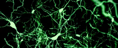
Tools that assist in the rapid visualization and quantification of neurons are necessary for monitoring neural health, and the effects of neural cell culture conditions, environmental neurotoxicants, biological modifiers, and drug compounds. Advances in high-quality microscopy and labeling techniques enable scientists to capture molecular interactions, protein expression patterns, and neural signaling pathways. By doing so, scientists can monitor neuronal subpopulation characterization (i.e., dopaminergic, cholinergic, GABAergic), neural cell culture conditions, and the effects of biological modifiers, drug compounds, and environmental neurotoxicants. Prominent and commonly utilized visualization techniques in the field include fluorescent labeling, endogenous labeling, imaging of 3D neural co-cultures, calcium imaging, and live-cell imaging, to name a few. The increasing availability of specialized equipment and technology has particularly allowed the proliferation of live-cell imaging techniques which require minimal training to obtain informative data and imagery on neurons and related processes. Microscopic analysis of neural networks is now more accessible than ever before and helps facilitate our understanding of the significant interplay between neurons, that receive and transmit chemical or electrical signals, and glial cells such as astrocytes and microglia, that serve as a supporting cast and play an information processing role complementary to the former. As various NDDs such as Alzheimer’s, Parkinson’s, and ALS are often associated with dysfunction or a deficiency in glial cells, the development and use of imaging technologies that help monitor these interactions are important in helping our understanding of NDD progression and development.
Thermo Fisher Scientific—High-content screening and compatible imaging reagents
Cellular imaging technologies from Thermo Fisher Scientific embody a broad technology platform for neuroscience research. For example, high-content imaging systems and associated compatible reagents from Thermo Fisher Scientific have helped neuroscientists to advance neurobiological research extending beyond the analysis of normal brain functioning to an intricate and detailed understanding of the cellular changes that characterize myriad NDDs.
The investigation of the complex and dynamic processes in living neurons requires the use of tools that can specifically target neurons. Furthermore, high-quality fluorescent labels are needed to specifically label neurons or visualize cellular processes to observe different neuron phenotypes. Products from Thermo Fisher Scientific encompass all major neural cell types and support a multitude of applications from basic research to therapeutic discovery, thus allowing scientists to streamline their neurobiology experiments and obtain relevant results. Reagents such as the PrestoBlue Cell Viability reagent facilitate measuring the metabolic activity of cells while dyes such as Calcein AM provide a useful measure of cell viability thanks to its dependency on enzymatic esterase activity and cell membrane integrity. Various cell function assays such as the CyQUANT Direct assay, the Ethidium Homodimer-1 (EthD-1) DNA stain, and neurite outgrowth staining kit from Thermo Fisher Scientific enable precise characterization and labeling of cellular structures and processes. Additional tools include antibody labeling kits, specifically, the Alexa FluorTM 647 Antibody Labeling kit and the pHrodoTM iFL Red Microscale Protein Labeling kit that have been used extensively in neuroscience research.
These efforts are further enhanced through High-Content Screening (HCS) or High-Content Analysis platforms (CX7 Pro platforms) from Thermo Fisher Scientific that are designed for exceptional single-cell analysis and ultra-fast time-to-data. These platforms provide for unbiased spontaneous phenotyping with intact, fixed, or live cells ranging from 2D monolayers to 3D spheroids while acquiring complex imagery and high-content data in seconds. The CellInsight CX7 Pro platform, live- and fixed-cell fluorescence labelling reagents, and HCS Studio software from Thermo Fisher Scientific combine multi-parameter fluorescence microscopy, via multiplexable fluorescent reagents, and integrated neuronal profiling bioapplication algorithms that identify and measure each cell within the fields of view. The high-throughput and ultra-fast capabilities of these platforms are made possible by conducting imaging and analysis in parallel and providing the fastest time-to-data possible. These capabilities are further enhanced by recent advances that Thermo Fisher Scientific has incorporated into its systems including laser-based focusing, confocal spinning disc performance, camera sensitivity, and dedicated neuronal profiling bioapplication processing. Consequently, Thermo Fisher Scientific has achieved accelerated throughput such that a 96-well plate of neurons can be fully scanned and quantified in less than four minutes (CX7 Pro).
Thermo Fisher Scientific in action
A representative example where Thermo Fisher Scientific has played a leading role in neuroscience research involves a study monitoring neurite morphology, outgrowth assessment, and synapse formation in primary neurons for drug screening and neurotoxicity assessments. Here, the CX7 HCS platform from Thermo Fisher Scientific was utilized for the quantitation of neuron morphology and synapses in vitro. As a result, this enabled the analysis and monitoring of neurite degeneration and neuron death in real time while allowing the measurement of related parameters in an automated process. The assay would go on to automatically identify the primary neuron cells, their morphology, synapses, and phenotypic features which are potential indicators for neuron development, differentiation, and neurotoxicity. The changes were then quantified in different conditions for different drug treatments thus providing a comprehensive evaluation of the development of neurotoxicity in neurites and synapses. In this manner, tools from Thermo Fisher Scientific have helped develop an assay that facilitates automation and streamlining of a laborious process in drug discovery screening and compound neurotoxicity assessment.
Similarly, HCS platforms from Thermo Fisher Scientific have found great application in other representative neuroscientific studies including the characterization of the anti-inflammatory properties of KLS-13019, a novel compound to treat and reverse chemotherapy-induced neuropathy. The CX5 platform from Thermo Fisher Scientific would be used exclusively to profile large numbers of neurons with the assay results revealing that KLS-13019 demonstrates anti-inflammatory properties. The study would solidify KLS-13019 as a potent neuroprotective and anti-inflammatory cannabinoid with great therapeutic potential for high efficacy treatment for neuropathic pain.
CX5’s counterpart in CX7 would be utilized in another study to determine alcohol toxicity in human neurons. Here again, the CX7 platform would assist in the profiling of a large population of neurons. This effort would be further supplemented using reagents and cell tracers from Thermo Fisher Scientific, such as the Alexa Fluor 488 conjugate, especially for neural cell characterization, to help differentiate neural progenitor cells from human dopaminergic neuron-containing cultures. This would help demonstrate the neurotoxic effects of chronic ethanol exposure, identify its modulating agents, and provide quantification of ethanol toxicity in live human neurons. Cell tracers from Thermo Fisher Scientific are non-toxic, stable probes that allow for cell movement and localization studies in live cells. Available in a range of fluorescent colors, cell tracers are an essential resource for neuroscientific studies.
Conclusion
The study of neurons is fundamental to our understanding of brain development and function. One of the major goals of neuroscience is to shed light on the dynamic processes and cellular changes behind devastating neurodegenerative diseases. Innovations in microscopic imaging technology have made available a variety of image-based assays that provide microscopic analysis of neurons and neural networks. Neuroscience imaging and analysis has never been more accessible than it is now.
Thermo Fisher Scientific offers end-to-end cell imaging products and resources. Take advantage of our expertise all found in one convenient place, with relevant tools, protocols, guides, insights and more, all at your fingertips. We are here for you.
Take advantage of our 40+ years of experience and expertise with end-to-end cell images products and resources, including relevant tools, protocols, selection guides, inspiration, and more at your fingertips. To learn more about cellular imaging and related technologies, visit the Thermo Fisher Scientific imaging resource center: www.thermofisher.com/cellularimaging.

